Review Article
Wifi and health: Perspectives and risks

Myriam Ben Salah1*, Hafedh Abdelmelek2 and Manef Abderraba1
1Institut Préparatoire aux Etudes Scientifiques et Techniques, Laboratoire Molécules Matériaux et Applications, la Marsa, Tunisia
2Faculte des Sciences de Bizerte, Laboratoire de Physiologie Intégrée, 7021Jarzouna, Tunisia
*Address for Correspondence: Myriam Ben Salah, Institut Préparatoire aux Etudes Scientifiques et Techniques, Laboratoire Molécules Matériaux et Applications, la Marsa, Tunisia, Email: [email protected]
Dates: Submitted: 23 June 2017; Approved: 09 October 2017; Published: 12 October 2017
How to cite this article: Salah MB, Abdelmelek H, Abderraba M. Wifi and health: Perspectives and risks. Ann Biomed Sci Eng. 2017; 1: 012-022. DOI: 10.29328/journal.abse.1001002
Copyright License: © 2017 Salah MB, et al. This is an open access article distributed under the Creative Commons Attribution License, which permits unrestricted use, distribution, and reproduction in any medium, provided the original work is properly cited.
Keywords: Radio frequency; Wireless; Biological effects; Oxidative stress

Abstract
Increased exposure to electromagnetic fields such as radio frequencies used by Wifi technology raise questions and concerns about their impact on health. For answer these questions, several scientific studies have carried out followed by results publication in prestigious scientific revues. Literature conducted on the effects of non-ionizing radiation and Wifi waves is vast and sometimes controversial. Epidemiological studies and the results of in vitro and in vivo experimental studies have showed the biological effects of electromagnetic field in different frequencies range. These effects caused disorders at the molecular and behavioral level. However, these studies were insufficient to confirm the directly related effects to the cause. Therefore, further research must be done to raise the controversy about the safety of wireless waves.
Introduction
The evolution of the information society and the use of new technologies make the magnetic field omnipresent in all areas of life. Increased exposure to this type of field raise questions and concerns about their impact on health. Invading magnetic fields application in industries (radars, telecommunications…) and medecine (IRM) increase general and professional exposure. Therefore, international regulation is required. In fact, associations in France and abroad are concerned about the health effects of electromagnetic radiation. These associations relay the concerns of citizens, alert the authorities and seize the courts to enforce the precautionary principle [1].within this context that the International Radiation Protection Committee (INC) has prepared recommendations on exposure limits for main types of non-ionizing radiation [2]. On the occasion of its 8th International Congress in Montreal (1992), the International Association of Radio Protection (IRPA) has set up a new independent scientific organization, International Commission for the Protection against Non-Ionizing Radiation Protection (ICNIRP) which takes over from the IRPA / INIRC. This commission’s role is to study the potential risks associated with various forms of non-ionizing radiation and studying the protection against radiation.
Many scientific studies have already been conducted on the health risks of radiation and electromagnetic waves. Barnothy [3], is the first to have conducted a review on the biological effects of electromagnetic fields followed by other works [4-8]. The laboratory of «physiologie intégrée» in «Faculté des Science de Bizerte » (Tunisia) was also interested in the effects of electromagnetic waves on biological systems. de Chater et al. [9], have showed that the static magnetic field induces oxidative stress and apoptosis in the thymus cells of pregnant rats. Moreover, Abdelmelek et al. [10], reported that the static magnetic field (128 mT, 5 days) induces hypoxia-like associated with sympathetic hyperactivity proving that the CM could be considered an environmental stress agent. Similarly, Amara et al. [11], showed that subchronic exposure to the CMS induced oxidative stress and DNA fragmentation after cell leakage zinc accompanied by a decrease in antioxidant activity of the enzyme system such as glutathione peroxidase and catalase. Other studies showed that subacute exposure to CMS (128 mT) caused metabolic disorders, hyperglycemia and insulin decrease like diabetes state [12-14]. It was able to extend the lifetime of free radicals and to change the some enzymes activity [15,16]. These biologic changes depend on waves/matter interaction at considered frequency. It is known that electromagnetic fields permanently immersed in a frequency ocean from low frequency (between 0 static fields and 100 KHz high-tension lines and domestic installation) to radio frequency (100 KHz and 300 KHz: television, microwave oven etc.). In this revew, we reported the biological effects of waves generated by WIFI technology. This is a wireless network technique for short-distance data exchange by microwave fields (RF) at 2.45 GHz. Since WiFi are electromagnetic waves, specifically radio frequency (RF), we thought that it too could induce metabolic disorders. There are few scientific studies on the physiological effects of WiFi as a recent technology. Most existing scientific studies support their hypotheses and thoughts on epidemiological studies on the effects of these RF on neurophysiology [1,17,18]. The biological effects of RF could be attributed to their thermal and non-thermal effects on health causing the appearance or development of pathologies. In fact, the RF could increase the production of free radicals and cause metabolic disorders such as diabetes [19,20].
Electromagnetic waves
For many years, Electromagnetic fields are a source of questions for the. Concerns were initially focused on power lines before moving recently to the antennas of mobile telephony. To answer these questions, thousands of scientific studies were launched, followed by the results published in prestigious scientific journals [5,6,16]. The use of electromagnetic fields has expanded to gradually gain industry, the medical field, homes and recently wireless transmission technologies that have invaded the High-Tech market (mobile phone, radio identification). Among these technologies, the WiFi is expanding rapidly and generates controversy. In fact, the use of the mobile phone generated a climate of suspicion about potential health risks of WiFi technology [21].
Definition of electromagnetic field
There is an electric field around every electrical installations caused by the electric charge transport due to the presence of an electric potential difference. The intensity of the created field depends on the voltage. Indeed, the magnetic field is the result from current produced by charges movement [22]. Therefore, Electromagnetic fields are the result of the combination of both electric and magnetic fields which move together with the speed of light. It are characterized by their frequency (hertz: Hz) and their wavelength [23].
Physical quantities and units
Magnetic field is characterized by the magnetic flux density (B), expressed in Tesla (T), and the magnetic field strength (H) expressed in Amperes per meter (A.m-1): B=μ*H
μ represents the physical constant of proportionality (magnetic permeability) (Institut National de Recherche et de Sécurité, 2011). Table 1 Quantities and SI units [24].
| Table 1: Quantities and SI units [24]. | |
| Quantity | Symbol and Unit |
| Electric field strength | E (V/m) |
| Electrical conductivity | σ (S/m) |
| Magnetic field strength | H (A/m) |
| Frequency | F or ν (Hz or s−1) |
| Magnetic flux density | B (T) |
| Permeability | µ (H/m) |
| Permittivity | Ε (F/m) |
| Power density | Ѕ (W/m2) |
| wave length | λ (m) |
| Specific Absorption Rate | DAS (W/kg) |
Sources of electromagnetic waves
Electromagnetic waves are emitted from two types of sources:
➢ Natural source
In our environment, we found Earth’s electromagnetic field generated by solar and atmospheric activities. This is a static magnetic field (25-65 microT). In fact, the orientation of the magnetic compass needle in the north-south direction is due to the Earth’s magnetic field. This field guides birds and fish during their migrations. In addition, the human body and specifically the heart and brain cells also produce low power electric and magnetic fields [25].
➢ Artificial sources
Electrical devices and modern communication generate electromagnetic waves such as microwave ovens, radios, television sets, magnetic resonance imaging, mobile phones and power lines [26,27]. Figure 1 electromagnetic spectrum and sources of non-ionizing and ionizing radiation (Federal office for radiation safety, Germany, 1999).
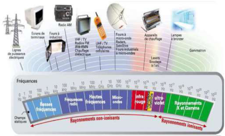
Figure 1: Electromagnetic spectrum and sources of non−ionizing and ionizing radiation (Federal office for radiation safety, Germany, 1999).
Electromagnetic spectrum
The electromagnetic spectrum is the decomposition of the electromagnetic radiation according to its different components (frequency, wavelength). Only a small portion of the spectrum is visible to the human eye called visible light. The properties of electromagnetic waves depend on their energy amount. X and gamma rays have the highest frequencies, followed by ultraviolet, visible light (red to violet), infrared, radio frequency or microwave s and extremely low frequencies [28].
Electromagnetic field properties
Magnetic waves are characterized by several physical parameters including:
• The electric field intensity (E),
• The magnetic field intensity (H),
• The magnetic flux density (B),
• The power density (S),
The variables related to perception and other indirect effects are contact current and, specific absorption (SA) (for pulsed fields). These quantities are used to define two limit values which are represented by:
• Basic restriction: limit values for exposure to electric, magnetic or electromagnetic at variable frequency. These values are established directly from the health effects [24].
• Reference levels: These levels are indicated for purposes of practical exposure assessment to determine if it is likely that the basic restrictions being exceeded. Some reference levels are derived from relevant basic restrictions using measurement techniques and/or calculation, and others are related to perception and to adverse indirect effects of exposure to electromagnetic fields [24].
Biological effects of electromagnetic field on human body
According to physical laws, the energy is as high as electromagnetic wave frequency is great. The effects of electromagnetic waves on the human body are highly dependent on proper mechanisms of action at each level of energy (Table 2).
| Table 2: Biological effect of electromagnetic field on human body [28]. | ||
| Radiation | Mecanism | Pathologic effects |
| X and gamma ray | Ionisation | Cancer |
| Ultraviolet | Chemical modifications | Skin cancer, skin aging, keratoconjunctivitis, Erythema. |
| Visible light | Photochemical réaction | Macular degeneration |
| Infrared | Surface heat | Cataract, retinal burn. |
| Microwaves and radiofrequencies | Deep heat | Temperature increase in Tissus |
| extremely low frequencies | Electric tension | Schock |
Biological effect of electromagnetic field on human body [28]
Tissue permeability to magnetic fields is the same as that of air. Consequently, the field within the tissues is identical to the external field. The humans and animals do not disturb this type of fields; their main interaction with the magnetic fields is expressed by Faraday’s law. Electric fields can also be induced by movement in a static magnetic field. Regarding the exposure of humans to magnetic fields, the main assessment criteria applied are:
• The most intense electric fields are induced in the most corpulent body;
• The induced electric field and associated current depend on the orientation of the magnetic field relative to the exposed body. The induced fields in the body have a maximum intensity when the field is perpendicular to the body;
• The distribution of the induced electric field is a function of the conductivity of the various organs and tissues [29].
The energy deposition in tissues depends on two major factors; the wavelength determines the reached depth, and radiation intensity. Objects effectively absorb the wave when they have approximately the same size as the wavelength. Thus, the infrared will be completely absorbed in the first few millimeters of the skin inducing stimulation of skin and sensation heat [28].
Effects of electromagnetic waves on biological systems
Literature conducted on the effects of non-ionizing radiation is large and sometimes controversial. Epidemiological studies and the results of in vitro and in vivo experimental studies carried out in recent years have highlighted the biological effects of electromagnetic field. These effects caused DNA and cell damage such as strand breaks of DNA, changes in the chromatin conformation, formation of micronucleus in different cell types, cell defects and apoptosis [30-32]. Other studies have found no effects observed following exposure to electromagnetic waves [33].
Various studies have shown that electromagnetic fields with low frequency and high frequency caused breaks in DNA strands. These results led the World Health Organization (WHO) in 2006 to give high priority to research into possible genetic modifications. Researchers have used the comet assay to observe any DNA damage induced by electromagnetic fields. Results showed that cells of human connective tissues exhibit a DNA strand breaks under the effect of low frequency electromagnetic fields alternately switched on and off at regular intervals [34].
Some results showed the existence of a relationship between exposure to electromagnetic fields and the increased incidence of the occurrence of some tumors types, particularly leukemia and brain cancer [35,36]. Several studies using different types of cancer cells showed contradictory effects on oxidative stress. Exposure of HL-60 cells to 100 mT for 13 min has no effect [37]. However, exposure for 2 hours to 6 mT increased oxidative stress in tumor cells U937 monocytic [38]. Reducing apoptosis may be a result of increased risk of carcinogenesis. Nuccitelli et al. [39], observed in tumor cells U937 monocytic a correlation between the reduction of apoptosis and modulation of membrane potential induced by exposure to 6 mT. These observations are supported by studies of Tenuzzo et al. [40], they exposed the cells to 6 mT up to 24 hours and noted a reduction of apoptosis and changes in the influx of free calcium. Abdelmelek et al. [10], reported that the static electromagnetic field (128T) increased the concentration of norepinephrine in skeletal muscle associated with sympathetic hyperactivity in rats. Chater et al. [9], showed that subacute exposure to static electromagnetic field stimulated the biosynthesis of plasma corticosterone and metallothionein in female rats and increased apoptosis. The mechanism of this stress effect induced by the electromagnetic field may be linked to oxidative stress [9]. There are many scientific data involving the electromagnetic field in the generation of free radicals such as superoxide anions in various cells (macrophages and monocytes)and organs (liver and kidney) [41-44]. Furthermore, several results showed that the magnetic field induced changes in enzyme activity, gene expression, changes in the structure and function of cell membrane and DNA damage [45-47]. In fact, previous studies showed that exposure to magnetic field increased DNA single-and double-strand breaks in rat brain [48]. A study on the treatment effect of zinc on oxidative stress induced by exposure to magnetic fields showed that the magnetic field (128 mT, 1 hour/day for 30 days) caused oxidative stress in the tissue et renal DNA. Indeed, it was able to of disturb the oxidant-antioxidant balance in different rat tissues. This imbalance was observed by a decrease in cytosolic GPx, CAT, and SOD activity in kidney and liver of rats. An increase in the level of MDA was also observed which could be explained by the lipid peroxidation [49].
Radiofrequencies
The spectrum of radio waves extends from 0.5 MHz in the AM radio band to about 30000 MHz in the radar band. Devices emitting radio frequencies have become ubiquitous in homes, offices and schools. Exposure to such a spectrum is quantified as radiofrequency energy flow per unit area (W/m²) [50]. Figure 2 band of radio waves [51]. Radiofrequency waves are long. Cell phones mainly use two frequency bands; 900 MHz (λ=33 cm) and 1.9 GHz (λ=16 cm). Therefore energy is deposited on a longer journey. According to the frequencies used by the various wireless technologies, the depth of penetration will reach a few centimeters [28]. Table 3 Penetration depth of radiofrequency into human tissues [28].
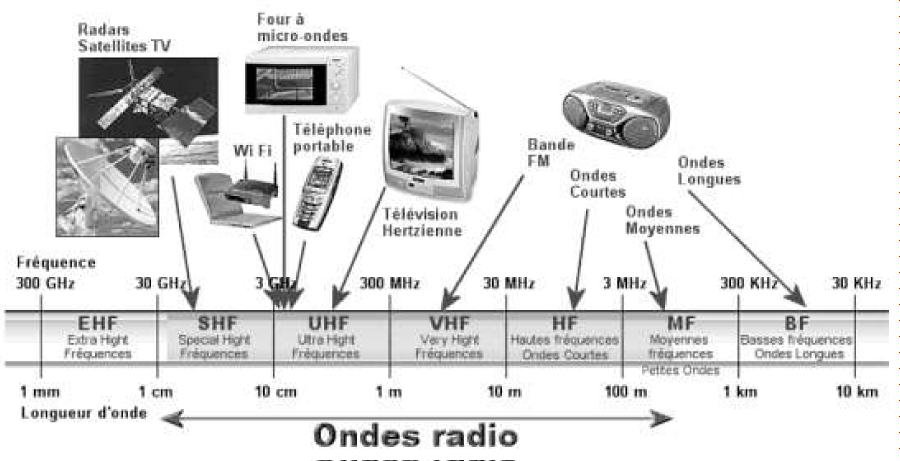
Figure 2: Band of radio waves [51].
| Table 3: Penetration depth of radiofrequency into human tissues [28]. | ||
| Wireless technologies | Frequency | Penetration deep |
| residential cordless phone systems | 5 GHz | <1 cm |
| Headset “Bluetooth”, Wifi | 2.4 GHz | 1 cm−2 cm |
| Cell phone | 1.9 GHz | 1 cm−3 cm |
| Cell phone | 900 MHz | 3 cm−5 cm |
| Radio waves (FM) | 100 MHz | >30 cm |
| IRM | 40 MHz, 100 MHz | >30 cm |
Characterization of wireless waves
Wireless waves or WiFi is a LAN wireless technology that uses radio frequencies as the carrier signal in order to extend the Ethernet network to a zone geographically distant. The connection between the different points is composed of an antenna for transmission and reception and a processing module for modulating and demodulating the signal [52].
Regarding the safety of emissions Wifi, two dangerous factors are advanced:
• Electromagnetic radiation inherent to the wireless technology;
• The value of used frequency which is around 2.4 GHz
In fact, the water resonance frequency which constitutes most of the human body is of 2.45 GHz. This frequency coincides with frequency spectrum used by WiFi which rising concerns among the general public [52].
The technical standard for transferring data by hertzian waves is IEEE 802.11 and its variants such as IEEE 802.11a, IEEE 802.11b. This standard is established by the organization of American standards IEEE-SA (Institute of Electrical and Electronics enginers-Standards Association). This standard describes the technical realization of the radio interface between the base station and mobile stations [53].
Signal propagation theory
The radio waves propagate in a straight line in several random directions which weaken with distance. When meeting an obstacle, part of its energy is absorbed and the other part continues to spread attenuated way. Radio waves can be reflected and diffracted when they encounter an angle [53].
Access points and bridges WiFi
An access point allows the interconnection of wireless equipment. Communication is limited to the signal coverage area that spreads 360° via Wifi antenna. We recommend a maximum of twenty clients per access point that must be placed in height to avoid environmental disturbances. The wireless bridge allows extending network to a remote geographical area. Indeed, the signal spreads several kilometers along the equipment. Therefore, the access point allows the use of inside network while the access bridge allows the use of the network outside and in open field [53].
Health regulations
The European Directive 2004/40/EC sets the minimum safety and health requirements for limiting workers’ exposure to electromagnetic fields. It is defined in terms of the values frequency of quantities relating to electric and / or magnetic beyond which preventive measures are to be implemented [29]. Table 4 Limiting public and workers’ exposure to electromagnetic fields [29].
| Table 4: Limiting public and workers' exposure to electromagnetic fields [29]. | ||||||||
| Standard | electric field strength | Magnetic field strength | Magnetic induction | Equivalent plane wave power density | ||||
| E (V/m) | H (A/m) | B (µT) | S eq (W/m2) | |||||
| Public | Workers | Public | Workers | Public | Workers | Public | Workers | |
| Bluetooth | 61 | 137 | 0.16 | 0.36 | 0.20 | 0.45 | 10 | 50 |
| DECT | 58 | 127 | 0.16 | 0.35 | 0.20 | 0.44 | 9.50 | 47.50 |
| Wifi | 61 | 137 | 0.16 | 0.36 | 0.20 | 0.45 | 10 | 50 |
| WiMax | 61 | 137 | 0.16 | 0.36 | 0.20 | 0.45 | 10 | 50 |
Biological effects of radiofrequency
Several studies showed that low-level RF energy could induce cancers such as leukemia, immunological disorders, deficiency in the blood-brain barrier, neurological anomalies such as headaches, disturbances in sleep and difficulty in concentration [54,55]. Exposure to low power RF can affect the cholinergic system. This sensibility could be due to a decrease of the intake of choline and activation of endogenous opioid neuroreceptors [56-58].
The causal chain that begins from RF exposure to death contains several steps. Each step may be the precursor to the next step. For interaction of radio frequencies with molecules, cellular structures or tissue, the transduction mechanism is the first crucial binding in the causal chain [50]. Figure 3 Radiofrequencies interaction with biological systems [50]. Radio frequencies and microwaves cause tissue heating. This thermal effect constitutes the standards basis of exposure for workers and the public. According to authors, the molecular damage caused by radio frequencies and microwaves activated the reaction of the cells. The natural defense mechanism was subsequently activated against this kind of stimulus. Such a reaction can be detected through the increase in reactive oxygen species or by the increase of stress proteins [59-61]. In addition, the conditions of exposure to mobile phones and DECT have caused real bioeffects in memory and brain of mice [62-65]. Figure 4 exposure and radiofrequencies effect at varied powers [66].
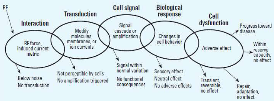
Figure 3: Radiofrequencies interaction with biological systems [50].
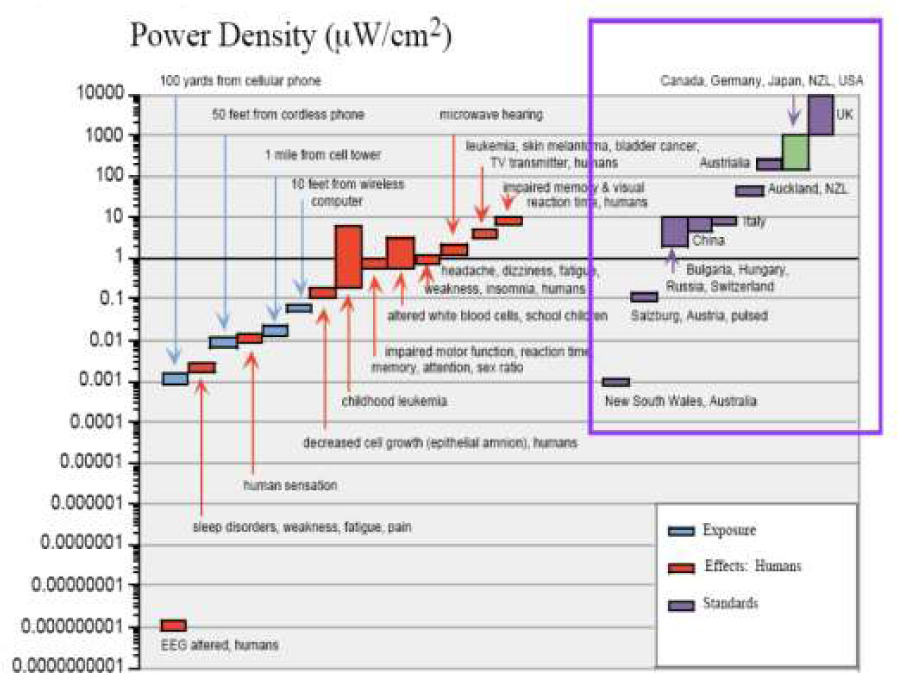
Figure 4: Exposure and radiofrequncies effect at varied powers [66].
Installation and use of Wifi in administrations, libraries and academic institutions are challenged. Indeed, some scientific studies showed that the WiFi are pulsed waves using the same frequency as that used in microwave ovens. Therefore, the risks should be evaluated both on their thermal effects (proportional to the power density) and their non-thermal effects in the medium and long term [1]. The power absorbed by biological tissue per unit time could be transform to stored energy which induced increase in temperature (hyperthermia) localized or in full body [29].
Consequently, effects of radiofrequencies could be:
• Thermogenic effects: electromagnetic waves are absorbed by living tissue and it degrades into heat causing hyperthermia. In the case of Bluetooth technology, WLAN and DECT, low power implementations could not disrupt the thermoregulatory mechanisms human body [29].
v• Non-thermogenic Effects: the electromagnetic waves could be causing some biological effects; cellular (cancer), endocrine, immunological, neurobiological effects and potentiating effects associated with other aggressive agents [17].
In addition, radio frequency interference with pacemakers is theoretically possible. This is an electromagnetic problem complicated by the biological environment (patient’s body). At a distance less than 10 cm between a radio and a pacemaker minor disturbances have been recorded [67,68].
Conclusion
In this work, we tried to characterize the electromagnetic field and specifically the RF and their interactions with biological systems. Several scientific studies showed existence radiation effect on health. However, these studies are insufficient to confirm the effects directly related to the cause. Thus, more advanced epidemiological research must be done to enhance or reduce the uncertainties raised about the health impact of electromagnetic waves.
References
- Esteves Sobral, Jordan PS. Teyssendier de Maistre (2009) Les Effets du Wifi sur la santé. Note de synthèse.
- UNEP/WHO/IRPA. United Nations Environment Programme/International Radiation. 1987.
- Barnothy MF. Biological effects of magnetic field. Plenum Press: New York Press. 1964.
- Adey WR. Biological effects of electromagnetic fields. J Cell Biochem. 1993; 51: 410-416. Ref.: https://goo.gl/YUw7nj
- Blank M. Biological effects of environmental electromagnetic fields: molecular mechanisms. Biosystems. 1995; 35: 175-178. Ref.: https://goo.gl/gZ6jo7
- Blank M. Electromagnetic fields: biological interactions and mechanisms. New York: American Chemical Society. 1995. Ref.: https://goo.gl/p84koe
- Binhi VN, Savin AV. Molecular gyroscopes and biological effects of weak extremely low frequency magnetic fields. Phys Rev E Stat Nonlin Soft Matter Phys. 2002; 65: 1-10. Ref.: https://goo.gl/jvm4Gp
- Starvroulakis P. Biological effects of electromagnetic fields. Berlin Heidelberg Springer-Varlag. Postgrad Med J. 2003; 80: 253-261. Ref.: https://goo.gl/sVsW4Q
- Chater S, Abdelmelek H, Couton D, Joulin V, Sakly M, et al. Subacute exposure to magnetic field induced apoptosis in thymus female rats. Pak J Med Sci. 2005; 21: 292-297. Ref.: https://goo.gl/s1Mcgd
- Abdelmelek H, Molnar S, Servais S, Cottet−Emard JM, Pequignot JM, et al. Skeletal muscle HSP72 and norepinephrine response to static magnetic field in rat. J Neural Transm. 2006; 113: 821-827. Ref.: https://goo.gl/KNg8Sh
- Amara S, Abdelmelek H, Abidi R, Sakly M, Ben Rhouma K. Zinc prevents hematological and biochemical alteration induced by static magnetic field in rats. Pharmacol Rep. 2006; 57: 616-622. Ref.: https://goo.gl/nC2tgc
- Chater S, Abdelmelek H, Pequignot JM, Sakly M, Rhouma KB. Effects of subacute exposure to static magnetic field on hematologic and biochemical parameters in pregnant rats. Electromagn Biol Med. 2006 ; 25: 135-144. Ref. : https://goo.gl/mAQTFT
- Elferchichi M, Mercier J, Coisy-Quivy M, Metz L, Lajoix AD, et al. Effects of exposure to a 128-mT static magnetic field on glucose and lipid metabolism in serum and skeletal muscle of rats. Arch Med Res. 2010; 41: 309-314. Ref. : https://goo.gl/Sa9ymB
- Lahbib A, Elferchichi M, Ghodbane S, Belguith H, Chater S, et al. Time-dependent effects of exposure to static magnetic field on glucose and lipid metabolism in rat. Gen Physiol Biophys. 2010; 29: 390-395. Ref.: https://goo.gl/WnKpJn
- Roy S, Noda Y, Eckert V, Traber MG, Mori A, et al. The phorbol 12-myristate 13-acetate (PMA)-induced oxidative burst in rat peritoneal neutrophils is increased by a 0.1 mT (60 Hz) magnetic field. FEBS Lett. 1995; 76: 164-166. Ref.: https://goo.gl/6DF13d
- Lacy-Hulbert A, Metcalfe JC, Hesketh R. Biological responses to electromagnetic fields. Faseb J. 1998; 12: 395-420. Ref.: https://goo.gl/Fg9AAE
- Crouzier D, Debouzy JC, Bourbon F, Collin A, Perrin A, et al. Neurophysiologic effects at low level 1.8 GHz radiofrequency field exposure: a multiparametric approach on freely moving rats. Pathologie Biologie. 2007; 55: 134-142. Ref.: https://goo.gl/UTTeYg
- Crouzier D, Testylier G, Perrin A, Debouzy JC. Which Neurophysiologic effects at low level 2.45 GHz exposure?. Pathologie Biologie. 2007; 55: 235-241. Ref.: https://goo.gl/yVctHF
- Akoev IG, Pashovkina MS, Dolgacheva LP, Semenova TP, Kalmykov V L. Enzymatic activity of some tissues and blood serum from animals and humans exposed to microwaves and hypothesis on the possible role of free radical processes in the nonlinear effects and modification of emotional behavior of animals. Radiat Biol Radioecol. 2002; 42: 332-330. Ref.: https://goo.gl/5grcK6
- Belpomme D, Irigaray P, Hardell L. Electromagnetic fields as cancer-causing agents. Environmental Research. 2008; 107: 289-290.
- Conseil Supérieur de la Santé (2008) Effets biologiques potentiels des micro-ondes modulées. Publication N°8194, 1
- Baptiste J (2001) Effets biologiques des ELF. Sciences et avenir : 87-88.
- Hée G, Méreau P, Dornier G (2002) Champs et ondes électromagnétique. Travail et sécurité
- Hygiène et sécurité du travail N°182, 1er trimestre (2001) Guide pour l’établissement de limites d’exposition aux champs électriques, magnétiques et électromagnétiques
- Feychting M. Health effects of static magnetic fields--a review of the epidemiological evidence, Prog Biophys Mol Biol, 2005; 87: 241-246. Ref.: https://goo.gl/TtJCmD
- Jelenkovic A, Janac B, Pesic V, Jovanovic DM, Vasiljevic I, et al. Effects of extremely low-frequency magnetic field in the brain of rats. Brain Research Bulletin. 2006; 68: 355-360. Ref.: https://goo.gl/SsL3v4
- Hashish AH, El-Missiry MA, Abdelkader HI, Abou-Saleh RH. Assessement of biological changes of continuous whole body exposure to static magnetic field and extremely low frequency electromagnetic fields in mice. Ecotoxicol Environ Saf. 2008; 71: 895-902. Ref.: https://goo.gl/XnfV65
- Plante M. Cellulaires et santé: êtes-vous sur la même longueur d’onde que vos patients?. Le Médecin du Québec. 2010; 45: 41-46.
- Institut National de Recherche et de Sécurité (INRS) (2012) Champs électromagnétiques: Les réseaux sans fil de proximité. ED 4207.
- Lai H. Singh NP. Single- and double-strand DNA breaks in rat brain cells after acute exposure to radiofrequency electromagnetic radiation. Int J Radiat Biol. 1996; 69: 513-521. Ref.: https://goo.gl/wUGG5y
- Lixia S, Yao K, Kaijun W, Deqiang L, Huajun H, et al. Effects of 1.8 GHz radiofrequency field on DNA damage and expression of heat shock protein 70 in human lens epithelial cells. Mutat Res. 2006; 602:135-142. Ref.: https://goo.gl/3VgLnx
- Zhao TY, Zou SP, Knapp PE. Exposure to cell phone radiation up-Regulates apoptosis genes in primary cultures of neurons and astrocytes. Neurosci Lett. 2007; 412: 34-38. Ref.: https://goo.gl/CyMVWH
- Belyaev IY, Koch CB, Terenius O, Roxström- Lindquist K, Malmgren LOH, et al. Exposure of rat brain to 915MHz GSM microwaves induces changes in gene expression but not double stranded DNA breaks or effects in chromatin conformation. Bioelectromagnetics. 2006; 27: 295-306. Ref.: https://goo.gl/PzZaYx
- Programme national de recherche PNR 57 Résultats du programme national de recherche sur les éventuels risques sanitaires émanant des rayonnements électromagnétiques. Rayonnement non ionisant. Environnement ET santé. 2011.
- Wertheimer N, Savitz DA, Leeper E. Childhood cancer in relation to indicators of magnetic fields from ground current sources. Bioelectromagnetics. 1995; 16: 86-96. Ref.: https://goo.gl/L5W3pp
- AldrichTE, Andrews KW, Liboff AR. Brain cancer risk and electromagnetic fields (EMFs) assessing the geomagnetic component. Arch Environ Health. 2001; 56: 314-319. Ref.: https://goo.gl/5ywgq1
- Rozanski C, Belton M, Prato FS, Carson JJ. Real-time measurement of cytosolic free calcium concentration in DEM-treated HL-60 cells during static magnetic field exposure and activation by ATP. Bioelectromagnetics. 2009; 30: 213-221. Ref.: https://goo.gl/ZF9RdD
- De Nicola M, Cordisco SC, Cerella MC, Albertini M, D'Alessio A, et al. Magnetic fields protect from apoptosis via redox alteration. Ann N Y Acad Sci. 2006; 1090: 59-68. Ref.: https://goo.gl/v5XkcA
- Nuccitelli S, Cerella C, Cordisco S, Albertini MC, Accorsi A, et al. Hyperpolarization of plasma membrane of tumor cells sensitive to antiapoptotic effects of magnetic fields. Ann NY Acad Sci. 2006; 1090: 217-225. Ref.: https://goo.gl/avn5tp
- Tenuzzo B, Vergallo C, Dini L. Effect of 6mT static magnetic field on the bcl-2, bax, p53 and hsp70 expression in freshly isolated and in vitro aged human lymphocytes. Tissue Cell. 2009; 41: 169-179. Ref.: https://goo.gl/P6xXkq
- Khadir R, Morgan JL, Murray JJ. Effects of 60 Hz magnetic field exposure on polymorphonuclear leukocyte activation. Biochem Biophys Acta. 1999; 1472: 359-367. Ref.: https://goo.gl/oFpwpB
- Kula B, Sobczak A, Kuska R. Effects of static and ELF magnetic fields on free-radical processes in rat liver and kidney. Electron Magnetobiol. 2000; 19: 99-105. Ref.: https://goo.gl/FWoA55
- Simko M, Droste S, Kriehuber R, Weiss DG. Stimulation of phagocytosis and free radical production in murine macrophages by 50 Hz electromagnetic field. Eur J Cell Biol. 2001; 80: 562-566. Ref.: https://goo.gl/YDdtZv
- Lupke M, Rollwitz J, Simko M. Cell activating capacity of 50 Hz magnetic fields to release reactive oxygen intermediates in human umbilical cord blood-derived monocytes and in Mono Mac 6 cells. Free Radic Res. 2004; 38: 985-993. Ref.: https://goo.gl/oPfU4k
- Savitz DA. Overview of occupational exposure to electric and magnetic fields and cancer, advancements in exposure assessment. Environ Health Perspect. 1995; 103: 69-74. Ref.: https://goo.gl/VrDCw6
- Lewy H, Massot O, Touitou Y. Magnetic field (50 Hz) increases N-acetyltransferase, hydroxyindole-O-methyltransferase activity and melatonin release through an indirect pathway. Int J Radiat Biol, 2003; 79: 431-435. Ref.: https://goo.gl/5SP48W
- Yokus B, Cakir DU, Akday MZ, Sert C, Mete N. Oxidative DNA damage in rats exposed to extremely low frequency electromagnetic fields. Free Radic Res. 2005; 39: 317-323. Ref.: https://goo.gl/zpBY8Y
- Lai H, Singh NP. Acute exposure to a 60Hz magnetic field increases DNA strand breaks in rat brain cells. Bioelectromagnetics. 2004; 18: 156-165.
- Amara S, Abdelmelek H, Garrel C, Guiraud P, Douki T, et al. Zinc supplementation ameliorates static magnetic field oxydative stress in rat tissues. Environmental Toxicology and Pharmacology. 2007; 23:193-197. Ref.: https://goo.gl/XVjwji
- Valberg PA, van Deventer TE, Repacholi MH. Workgroup Report, Base Stations and Wireless Networks-Radiofrequency (RF) Exposures and Health Consequences. Environmental Health Perspectives, 2007; 115: 416-424. Ref.: https://goo.gl/VbzGE8
- Micallef N, Vallé S, Woringer M. Effets des ondes électromagnétiques sur le vivant Culture libre. sciences ouverte. 2008.
- Brunel JL. Les risques liés au WiFi. Observatoire Académique de la Sécurité Informatique: études/actualités. 2004.
- Mokdad M. Wi-Fi et son usage Contexte et implication. CIW, 2007, Le WI-FI. 2009.
- Hardell L, Mild KH. Re: “cellular telephone use and risk of acoustic neuroma” (author reply 4-5). Am J Epidemiol. 2004; 160: 923-926. Ref.: https://goo.gl/VQLhBX
- Lonn S, Ahlbom A, Hall P, Feychting M. Mobile phone use and the risk of acoustic neuroma. Epidemiology. 2004; 15: 653-659. Ref.: https://goo.gl/4G7dSL
- Lai H, Horita A, Chou CK, Guy AW. Effects of low-level microwave irradiation on hippocampal and frontal cortical choline uptake are classically conditionable. Pharmacol Biochem Behav. 1987; 27: 635-639. Ref.: https://goo.gl/YnQ9H9
- Lai H, Carino MA, Horita A, Guy AW. Low-level microwave irradiation and central cholinergic systems. Pharmacol Biochem Behav. 1989; 33: 131-138. Ref.: https://goo.gl/ApRU22
- Lai H, Carino MA, Wen YF, Horita A, Guy AW. Naltrexone pretreatment blocks microwave induced changes in central cholinergic receptors. Bioelectromagnetics. 1991; 12: 27-33. Ref.: https://goo.gl/pLmLy2
- Kwee S, Raskmark P, Velizarov S. Changes in cellular proteins due to environmental nonionizing radiation. I. Heat-shock proteins. Electro Magnetobiol. 2001; 20: 141-152. Ref.: https://goo.gl/JtCejg
- Friedman J, Kraus S, Hauptman Y, Schiff Y, Seger R. Mechanism of short-term ERK activation by electromagnetic fields at mobile phone frequencies. Biochem J. 2007; 405:559-568. Ref.: https://goo.gl/5ShXWY
- Blank M, Goodman R. Electromagnetic fields stress living cells. Pathophysiology. 2009; 16: 71-78. Ref.: https://goo.gl/29j8ZU
- Fragopoulou AF, Margaritis LH. Is cognitive function affected by mobile phone radiation exposure? In: L. Giuliani, M. Soffritti, eds. Non Thermal Effects and Mechanisms of interaction between electromagnetic fields and living matter. European Journal of Oncology-Library. 2010; 5: 261-273. Ref.: https://goo.gl/tdGN2r
- Fragopoulou A, Grigoriev Y, Johansson O, Margaritis LH, Morgan L, et al. Scientific panel on electromagnetic field health risks-consensus points, recommendations and rationales. Reviews on Environmental Health. 2010; 25: 307-317. Ref.: https://goo.gl/EdiKB8
- Ntzouni MP, Stamatakis A, Stylianopoulou F, Margaritis LH. Short term memory in mice is affected by mobile phone radiation. Pathophysiology. 2011; 18: 193-199. Ref.: https://goo.gl/9LgK8R
- Ntzouni MP, Skouroliakou A, Kostomitsopoulos N, Margaritis LH. Transient and cumulative memory impairements induced by GSM 1.8 GHz cell phone signal in a mouse model. Electromagn Biol Med. 2013; 32: 95-120. Ref.: https://goo.gl/Un2ZdW
- Firstenberg A. Radio Wave Packet. President, Cellular Phone Taskforce. 2001. Ref.: https://goo.gl/8mH4M4
- Butrous GS, Male JC, Webber R S, Barton DG, Meldrum SJ, et al. The effect of power frequency high intensity electric fields on implanted cardiac Pacemakers. Pacing Clin Electrophysiol. 1983; 6: 1282-1292. Ref.: https://goo.gl/Zi5BKG
- Kainz W, Casamento JP, Ruggera P S, Chan DD, Witters DM. Implantable cardiac pacemaker electromagnetic compatibility testing in a novel security system simulator. IEEE Trans Biomed Eng. 2005; 52: 520-530. Ref.: https://goo.gl/AbaWwj
- Elferchichi M, Mercier J, Abdelmelek H, Sakly M, Lambert K, et al. Effects of exposure to a 128-mT static magnetic field on glucose and lipid metabolism in serum and skeletal muscle of rats. Arch Med Res. 2010; 41: 309-314. Ref.: https://goo.gl/bNTFkK