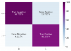
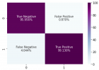
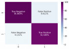
Figure 4
Applications of convolutional neural networks in chest X-ray analyses for the detection of COVID-19
Patrick Ting*, Anish Kasam and Kevin Lan
Published: 20 January, 2022 | Volume 6 - Issue 1 | Pages: 001-007

Figure 4:
Visualization of the ResNet50 neural network and its 50 layers. The convolutions are separated from the fully connected neural network and thus the model does not need to iterate through every neuron in the network [14].
Read Full Article HTML DOI: 10.29328/journal.abse.1001015 Cite this Article Read Full Article PDF
More Images
Similar Articles
-
A review article on artificial intelligenceNagendraswamy C*,Amogh Salis. A review article on artificial intelligence. . 2021 doi: 10.29328/journal.abse.1001012; 5: 013-014
-
Applications of convolutional neural networks in chest X-ray analyses for the detection of COVID-19Patrick Ting*,Anish Kasam,Kevin Lan. Applications of convolutional neural networks in chest X-ray analyses for the detection of COVID-19. . 2022 doi: 10.29328/journal.abse.1001015; 6: 001-007
-
Feasibility study of magnetic sensing for detecting single-neuron action potentialsDenis Tonini,Kai Wu,Renata Saha,Jian-Ping Wang*. Feasibility study of magnetic sensing for detecting single-neuron action potentials. . 2022 doi: 10.29328/journal.abse.1001018; 6: 019-029
-
Artificial awareness, as an innovative learning method and its application in science and technologyAdamski Adam*,Adamska Julia. Artificial awareness, as an innovative learning method and its application in science and technology. . 2023 doi: 10.29328/journal.abse.1001020; 7: 012-019
Recently Viewed
-
Effectiveness of prenatal intensive counselling on knowledge, attitude and acceptance of post placental intrauterine contraceptive device among mothersM Shanthini,Manjubala Dash*,A Felicia Chitra,S Jayanthi,P Sujatha. Effectiveness of prenatal intensive counselling on knowledge, attitude and acceptance of post placental intrauterine contraceptive device among mothers. Clin J Obstet Gynecol. 2020: doi: 10.29328/journal.cjog.1001044; 3: 021-025
-
Timely initiation of breastfeeding and associated factors among mothers who have infants less than six months of age in Gunchire Town, Southern Ethiopia 2019Ephrem Yohannes*,Tsegaye Tesfaye. Timely initiation of breastfeeding and associated factors among mothers who have infants less than six months of age in Gunchire Town, Southern Ethiopia 2019. Clin J Obstet Gynecol. 2020: doi: 10.29328/journal.cjog.1001045; 3: 026-032
-
Amenorrhea-An abnormal cessation of normal menstrual cycleNida Tabassum Khan*,Namra Jameel . Amenorrhea-An abnormal cessation of normal menstrual cycle. Clin J Obstet Gynecol. 2020: doi: 10.29328/journal.cjog.1001046; 3: 033-036
-
Maternal, neonatal and children´s health in Sub-Saharan East AfricaJosef Donát*. Maternal, neonatal and children´s health in Sub-Saharan East Africa. Clin J Obstet Gynecol. 2020: doi: 10.29328/journal.cjog.1001049; 3: 043-045
-
Determinants of neonatal near miss among neonates admitted to Ambo University Referral Hospital and Ambo General Hospital, Ethiopia, 2019Ephrem Yohannes*,Nega Assefa,Yadeta Dessie. Determinants of neonatal near miss among neonates admitted to Ambo University Referral Hospital and Ambo General Hospital, Ethiopia, 2019. Clin J Obstet Gynecol. 2020: doi: 10.29328/journal.cjog.1001050; 3: 046-053
Most Viewed
-
Feasibility study of magnetic sensing for detecting single-neuron action potentialsDenis Tonini,Kai Wu,Renata Saha,Jian-Ping Wang*. Feasibility study of magnetic sensing for detecting single-neuron action potentials. Ann Biomed Sci Eng. 2022 doi: 10.29328/journal.abse.1001018; 6: 019-029
-
Evaluation of In vitro and Ex vivo Models for Studying the Effectiveness of Vaginal Drug Systems in Controlling Microbe Infections: A Systematic ReviewMohammad Hossein Karami*, Majid Abdouss*, Mandana Karami. Evaluation of In vitro and Ex vivo Models for Studying the Effectiveness of Vaginal Drug Systems in Controlling Microbe Infections: A Systematic Review. Clin J Obstet Gynecol. 2023 doi: 10.29328/journal.cjog.1001151; 6: 201-215
-
Causal Link between Human Blood Metabolites and Asthma: An Investigation Using Mendelian RandomizationYong-Qing Zhu, Xiao-Yan Meng, Jing-Hua Yang*. Causal Link between Human Blood Metabolites and Asthma: An Investigation Using Mendelian Randomization. Arch Asthma Allergy Immunol. 2023 doi: 10.29328/journal.aaai.1001032; 7: 012-022
-
Impact of Latex Sensitization on Asthma and Rhinitis Progression: A Study at Abidjan-Cocody University Hospital - Côte d’Ivoire (Progression of Asthma and Rhinitis related to Latex Sensitization)Dasse Sery Romuald*, KL Siransy, N Koffi, RO Yeboah, EK Nguessan, HA Adou, VP Goran-Kouacou, AU Assi, JY Seri, S Moussa, D Oura, CL Memel, H Koya, E Atoukoula. Impact of Latex Sensitization on Asthma and Rhinitis Progression: A Study at Abidjan-Cocody University Hospital - Côte d’Ivoire (Progression of Asthma and Rhinitis related to Latex Sensitization). Arch Asthma Allergy Immunol. 2024 doi: 10.29328/journal.aaai.1001035; 8: 007-012
-
An algorithm to safely manage oral food challenge in an office-based setting for children with multiple food allergiesNathalie Cottel,Aïcha Dieme,Véronique Orcel,Yannick Chantran,Mélisande Bourgoin-Heck,Jocelyne Just. An algorithm to safely manage oral food challenge in an office-based setting for children with multiple food allergies. Arch Asthma Allergy Immunol. 2021 doi: 10.29328/journal.aaai.1001027; 5: 030-037

If you are already a member of our network and need to keep track of any developments regarding a question you have already submitted, click "take me to my Query."


